Your address will show here +12 34 56 78
Deep Learning, Medical Imaging

For prostate cancer patients, large organ deformations occurring between radiotherapy treatment sessions create uncertainty about the dose delivered to the tumor and surrounding healthy organs. Segmenting those regions on cone beam CT (CBCT) scans acquired on treatment day would reduce such uncertainties. In our latest work, a 3D U-net deep-learning architecture was trained to segment bladder, rectum and prostate on CBCT scans. Due to the scarcity of contoured CBCT scans, the training set was augmented with CT scans already contoured in the current clinical workflow. Our network was then tested on CBCT scans. The segmentation accuracy (as measured by the Dice similarity coefficient) increased significantly with the number of CBCT and CT scans in the training set, reaching a performance 10% better than conventional approaches based on deformable image registration between planning CT and treatment CBCT scans. Interestingly, adding CT scans to the CBCT training set allowed maintaining high performance, while halving the number of CBCT scans. Hence, our work showed that although CBCT scans included artifacts, cross-domain augmentation of the training set was effective and could rely on large datasets available for planning CT scans.

Collaborations
CHU-UCL-Namur, Belgium
CHU-Charleroi Hôpital André Vésale, Belgium

Links
Publisher: https://www.mdpi.com/2076-3417/10/3/1154
Github: https://github.com/eliottbrion/pelvis_segmentation

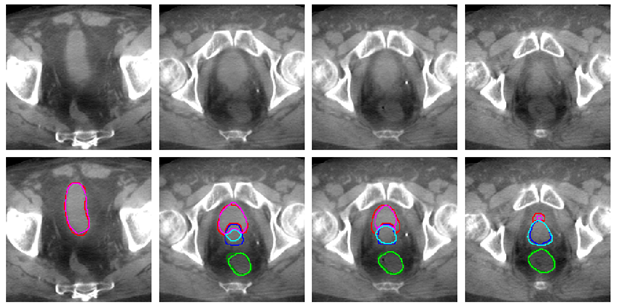
In this use case, the prostate is irradiated while minimizing the dose delivered to the healthy bladder and rectum. The image shows an example of automated bladder, rectum and prostate tracking on the CBCT scan acquired before each treatment cession. On this image, each column corresponds to a slice of the same CBCT scan. Dark colors represent the segmentation drawn manually by a human expert, while light colors show our algorithm segmentation. The automated bladder, in pink, rectum, in light green and prostate, in light blue, are close to human expert delineations. A further step will be to highlight these imporvements by showing better tumor coverage and reduction in the doses delivered to the healthy organs that it allows.

0

Deep Learning, Medical Imaging

TCLearn is a scalable method enabling multiple partners to participate together in distributed learning. Each new iteration of the model is first validated through federated byzantine agreement to guarantee the quality of the resulting model before being recorded in a blockchain. TCLearn preserves the privacy of both the data and the model while ensuring its optimal efficiency.

Link: 10.1109/ACCESS.2019.2959220

Full abstract

Distributed learning across coalitions is becoming popular for multi-centric implementation of deep learning models. However, the level of trust between the members of a coalition can vary and requires different security architectures. Privacy of the training data has been largely described in distributed learning. In this paper, we present a scalable security architecture providing additional features such as validation on the sources quality, confidentiality on the model within a trusted coalition or confidentiality among untrusted partners inside the coalition. More specifically, we propose solutions that guarantee preservation not only of data privacy but also of data quality, enforce a trustworthy sequence of iterative learning, and that lead to equitable sharing of the learned model among the coalition’s members. We give an example of its deployment in the case of the distributed optimization of a deep learning convolutional neural network trained on medical images.

Distributed learning across coalitions is becoming popular for multi-centric implementation of deep learning models. However, the level of trust between the members of a coalition can vary and requires different security architectures. Privacy of the training data has been largely described in distributed learning. In this paper, we present a scalable security architecture providing additional features such as validation on the sources quality, confidentiality on the model within a trusted coalition or confidentiality among untrusted partners inside the coalition. More specifically, we propose solutions that guarantee preservation not only of data privacy but also of data quality, enforce a trustworthy sequence of iterative learning, and that lead to equitable sharing of the learned model among the coalition’s members. We give an example of its deployment in the case of the distributed optimization of a deep learning convolutional neural network trained on medical images.

0

Medical Imaging

Because it is sensitive to the microscopic displacements of water molecules diffusing in the tissues, diffusion-weighted magnetic resonance imaging (DW-MRI) has become the modality of choice to study the anatomy of the white matter at the micrometer level, with applications in the diagnosis and treatment of many neurological disorders and brain injuries. Our new approach at the intersection of physical modeling, numerical simulations and signal processing proposes a way to bridge the gap between the native millimeter resolution of MRI and neuroanatomical indices at the micrometer scale.

We use specific MRI acquisitions known as diffusion-weighted MRI which by design are sensitive to the micrometer-scale diffusing motion of water molecules in the tissues at body temperature. In traditional approaches, forward mathematical models based on analytical formulas are devised to link the geometry of white matter tissue, the diffusion of water molecules in that geometric arrangement and the physics of MRI acquisitions. The free parameters of such a forward model are then adjusted to new MRI measurements by minimizing a cost function, providing indices of the white matter microstructure locally in each voxel. Such indices usually reflect the morphometry of the axons and glial cells making up the white matter.
In our latest work, we presented a forward model in which the MRI signal is not modeled analytically but instead computed by numerical simulations of the random walk of water in a 3D mesh representing the tissue. Such simulations, known as Monte Carlo simulations, provide signals that are more physically accurate than most traditional closed-form analytical models used in the literature. Due to their long computation times, their use has been mainly restricted to validation rather than directly in a model of the signal. In addition, being numerical results rather than mathematical formulas, they do not easily lend themselves to continuous optimization frameworks for parameter estimation. To circumvent these difficulties, we made use of combinatorial and convex optimization on a presimulated collection of MRI signals, or fingerprints, wherein each fingerprint uniquely characterizes a typical microstructural configuration.
On a rat model of spinal cord injury, we showed that the microstructural indices obtained with our model based on in vivo, non-invasive MRI were consistent with histological observations performed after sacrificing the animal. In particular, our model was able to detect axonal loss in the injured part of the spinal cord. The method was then applied on in vivo human brain data. Indices of axon diameter and density in specific white matter pathways were found to agree with previous histological observations in the human brain.
Future work will focus on using 3D geometries which more closely mimic white matter tissue in our presimulated dictionary of fingerprints. The method will also be applied on existing clinical datasets. This work encourages the use of computational models of the diffusion signal and gets us a step closer to quantitative characterization of the white matter microstructure in vivo.
Collaborations
Signal Processing Lab (LTS5), Ecole polytechnique fédérale de Lausanne, Switzerland
Computational Radiology Laboratory, Boston Children’s Hospital, Harvard Medical School, Boston, USA
Institute of NeuroScience, Université catholique de Louvain (Mont-Godinne), Belgium

Links
Publisher: https://www.sciencedirect.com/science/article/pii/S1053811918319487
Twitter (@MaximeTaquet): https://twitter.com/MaximeTaquet/status/1048534818831646722

0

Medical Imaging
ImagX.R, the research group on image guided proton therapy, has the pleasure to announce the PhD defence of Sylvain Deffet. He will present his research on proton radiography and discuss how this method could improve treatment outcome in proton therapy.

Proton therapy is an advanced form of radiation therapy which is increasingly used worldwide. Unlike photons, protons deliver a sharp dose at a precise location corresponding to their range in the patient. Thanks to this physical property, proton therapy has the potential to spare healthy tissues better than conventional radiation therapy. However, a consequence of this dosimetric property is that the range of the protons inside the patient must be accurately predicted to deliver the dose as planned. Unfortunately, several uncertainties arising during treatment planning may significantly impact the range of the protons and hence jeopardize dose conformity. To better quantify and potentially reduce the uncertainties, the implementation of imaging techniques that would provide a direct information on the energy reduction of protons in the patient is highly desirable. In the present thesis, such a method is introduced: proton radiography. The system that we propose relies on the use of protons having an energy high enough for them to traverse the patient and stop inside a detector which measures their residual range. By comparing measured and predicted residual ranges, it is possible to quantify range uncertainties in the patient in clinical conditions.

Although the principle of proton radiography is relatively straightforward, a clinical implementation of such a measurement device is a complex issue. This thesis aims at developing a proton radiography system and also at conceiving an acquisition process that could be conveniently implemented in clinics. The methodologies to correctly take advantage of proton radiography measurements are also discussed, in order to further optimize the dose delivered to the patient and improve de facto the treatment outcome.
0